
The term “scoliosis” originates from the Greek word meaning “curved”, which accurately reflects the nature of the condition. Scoliosis is considered one of the most complex challenges in modern orthopedics. It is characterized by a combination of typical functional, morphological, and radiological changes in the spine, rib cage, pelvis, and the positioning of internal organs.
According to various studies, the prevalence of scoliosis among children ranges from 5% to 14%. Based on epidemiological research conducted by the National Institute for Child and Mother Health in Moldova (2010), the prevalence of scoliosis in the Republic of Moldova reaches 5.2% of all musculoskeletal disorders.
Some researchers report that the average life expectancy of untreated patients with severe forms of scoliosis is only 35–40 years.
Definition
Scoliosis (scoliotic disease) is a lateral curvature of the spine accompanied by vertebral rotation. A distinctive feature of scoliosis is its progressive nature, which is closely linked to the child’s age and growth. Even in its early stages, true scoliosis involves structural spinal deformities that persist regardless of body position or physical load.
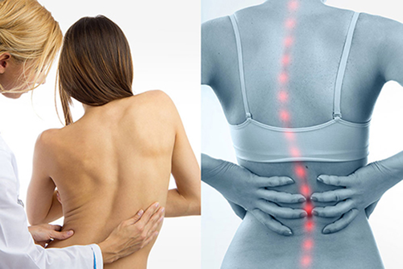
Diagnosis of Scoliosis
Early diagnosis plays a crucial role in the successful treatment of scoliosis and in preventing severe progression. Diagnostic steps typically include:
1. Clinical Examination
- Postural asymmetry (shoulders, scapulae, waistline, pelvis)
- Adam’s Forward Bend Test to detect spinal rotation and rib hump
- Palpation of the spinal column for curvature or rigidity
- Neurological assessment to rule out secondary causes
2. Radiographic Imaging
- X-rays (anteroposterior and lateral views) are the gold standard
- Cobb angle measurement determines the degree of spinal curvature:
- Mild: 10–20°
- Moderate: 20–40°
- Severe: >40°
- Spinal rotation and vertebral wedging may also be visualized
3. Advanced Imaging (if needed)
- MRI: indicated in cases of rapid progression, neurological symptoms, or congenital anomalies
- CT scan: used for detailed bone analysis or pre-surgical planning
4. Functional Tests
- Assessment of spinal flexibility and posture
- Gait analysis and evaluation of muscle tone
- Respiratory function tests in severe thoracic scoliosis
5. Monitoring Progression
Regular monitoring is essential, especially during growth spurts. X-rays are repeated every 6–12 months, depending on the severity and progression rate.
11 лет
13 лет
15 лет
Recognition of scoliosis is extremely important at the early stages of its development, because only timely started treatment can prevent the progression of the curvature. It is important to know at what age the spinal deformity was noticed and how it progressed.
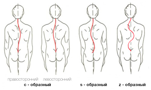
In patients with scoliosis, pelvic tilt, lateral deviation of the torso, and lateral curvature of the spine are observed. When scoliosis is present, one shoulder is higher than the other, which can be easily determined by the level of the scapulae and clavicles. The scapula on the concave side of the curvature is closer to the spinous processes than on the convex side. The distance from the top of the spinous process of the seventh cervical vertebra (C7) to the scapular angle is smaller on the convex side of the curvature than on the concave side. The degree of stability of the deformity is assessed by gently pulling the head.
X-rays must be taken in two projections, both lying down and standing up. The severity of scoliosis is characterized by the degree of changes. Patients with congenital scoliosis often have changes in the cardiovascular and respiratory systems. If scoliosis develops in the thoracic spine, the deformed vertebrae pull the attached ribs along with them, causing deformation of the chest and development of a rib hump.
An orthopedic specialist’s focus during scoliosis treatment should be on identifying and correcting the primary curve, which is the main task. The longer scoliosis exists, the more stable and fixed it becomes, and the harder it is to correct the deformity.
The progression of scoliosis depends on the patient’s age, type, degree of curvature, and cause. The greatest progression is observed during the child’s rapid growth phase and usually stops when growth ceases. Therefore, the earlier the disease starts, the higher the risk of deformity progression; the later it starts, the less chance there is of significant scoliosis development.
Scoliosis progression continues with the child’s growth and reaches its maximum during puberty — at ages 11–13 in girls and 14–16 in boys. From this age, the progression rate gradually decreases and stops by the end of skeletal growth, approximately between 17 and 20 years.
Scoliosis is a disease of the growing organism!
The main cause of deformity progression is the asymmetric growth of the vertebra.
The increase in scoliotic deformity in adults is caused by age-related changes in the bone tissue and degenerative changes in the intervertebral discs. Essentially, this is the “settling” or collapse of the deformed spine.
Classification of Scoliosis According to V. D. Chaklin:
- Grade I: Characterized by a lateral deviation up to 10° and an initial degree of torsion, detectable by X-ray.
- Grade II: Manifested by torsion and the presence of compensatory curves. Clinically, a muscular ridge and a small rib hump caused by vertebral torsion are observed. Lateral deviation ranges from 11° to 24°.
- Grade III: More pronounced deformity with lateral deviation between 25° and 40°, accompanied by a large rib hump. Radiologically, wedge-shaped vertebrae are seen at the apex of the curvature.
- Grade IV: Severe trunk deformity characterized by thoracic kyphoscoliosis, pronounced rib hump, pelvic deformation, and trunk deviation. The primary curve angle exceeds 41°.
Risser Sign (Risser Test)
The Risser sign is an objective clinical indicator used to assess the growth potential of the spine and to estimate skeletal maturity. It is based on the appearance of ossification centers on the iliac crest, visible on pelvic X-rays. These ossification points are among the last growth plates to close in the human body.
On an X-ray, these ossification zones look like a faint “cloud” floating above the iliac crest. When this “cloud” disappears—meaning it has fused with the bone—it indicates that skeletal growth has ended. This usually occurs around ages 16-18 in boys and slightly earlier in girls, before age 16.
Ossification begins at the anterior superior iliac spine and progresses posteriorly. The iliac crest is divided into four parts, and the degree of skeletal maturity is determined by how many of these parts are ossified:
| Risser Stage | Description |
| 0 | Apophysis not visible |
| 1 | Beginning of lateral ossification |
| 2 | Ossification of half the iliac crest |
| 3 | Beginning of fusion of the apophysis with iliac wing |
| 4 | Half fusion of the apophysis with iliac wing |
| 5 | Complete fusion of the apophysis with iliac wing |
- Stage 3 means approximately 75% ossification of the iliac crest.
- Stage 4 means all four parts are ossified.
- Stage 5 means complete fusion of the ossified apophysis with the iliac bone.
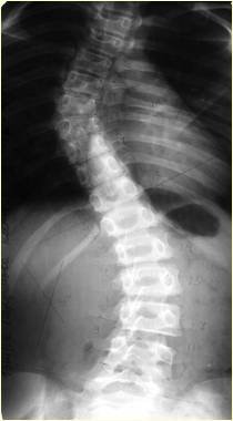
Cobb Method
Among the many methods for measuring the curvature angle of scoliosis, the Cobb method is the most widely used. It is based on determining the angle formed by the end vertebrae of the curvature arc.
On a frontal X-ray, lines are drawn along the upper endplate of the uppermost vertebra and the lower endplate of the lowermost vertebra of the scoliotic curve. Then, perpendicular lines are drawn from these two lines, and the angle formed between these perpendiculars is measured in degrees. This angle represents the degree of spinal curvature.
- For small curvatures, an additional angle between the perpendicular lines to these reference lines may be measured.
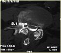
- If necessary, computed tomography (CT) scans can be performed to get more detailed imaging.
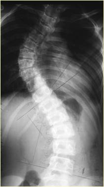
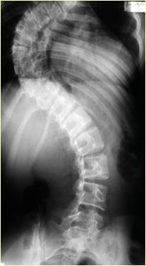
Example:
X-ray and CT of a 16-year-old patient diagnosed with idiopathic scoliosis, Grade 4, with a Cobb angle of 125 degrees.
Scoliosis Severity Classification by Cobb Angle:
Grade 4: Very severe scoliosis
Grade 1: Mild scoliosis
Grade 2: Moderate scoliosis
Grade 3: Severe scoliosis